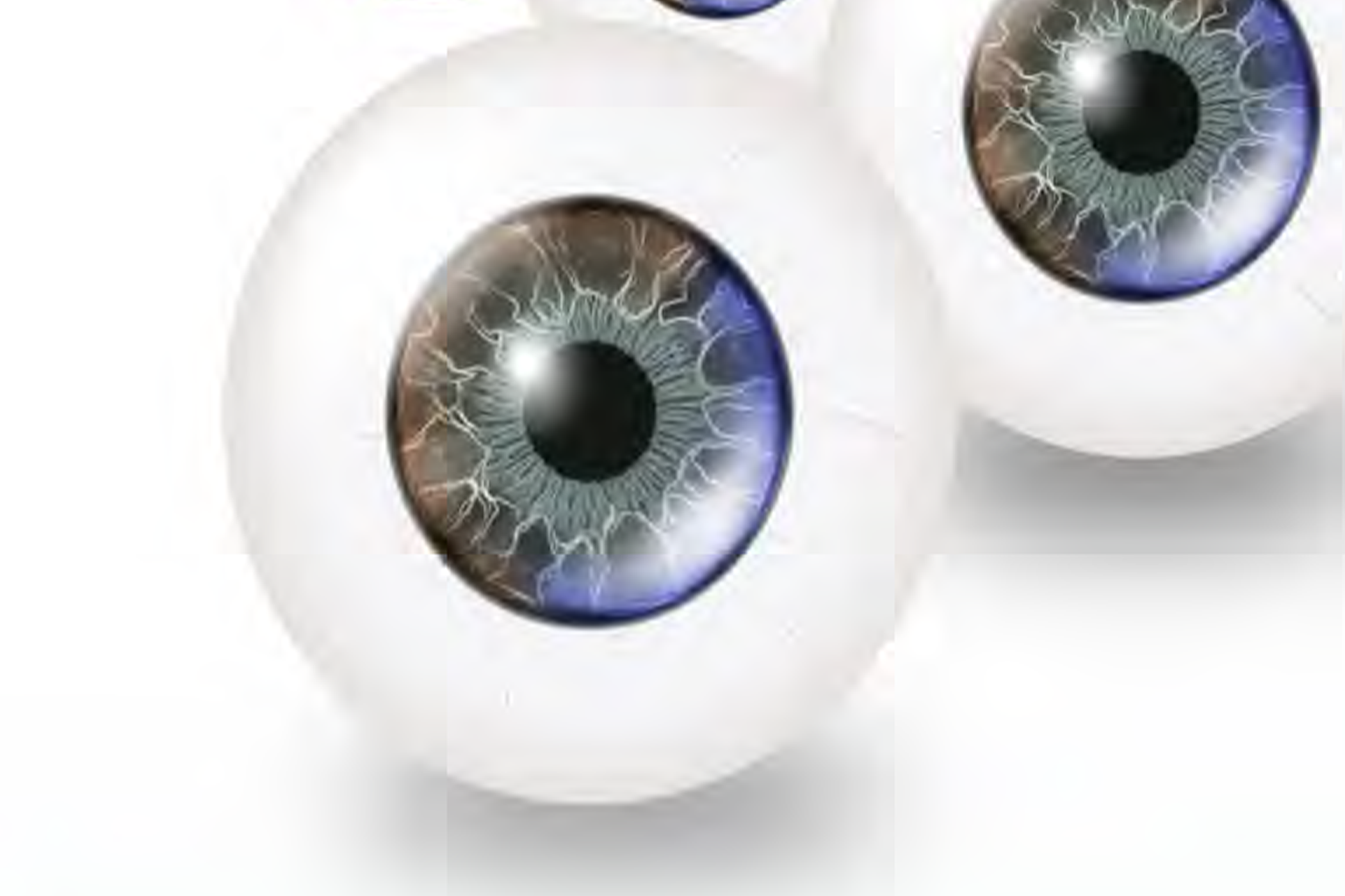
Artificial Eye

Detail
In medical training, the Department of Ophthalmology for diagnosing ocular complications using high-frequency ultrasound (ocular ultrasonography) has been limited to theoretical instruction. This lack of practical training can result in diagnostic errors when dealing with real patients. Accurate diagnosis, especially for localizing masses or foreign objects within the eye, requires continuous practice to identify abnormalities or foreign objects accurately and without confusion. To address this, researchers have developed artificial eyes or ocular models to enhance the training process. These models allow for effective learning and practice in diagnosing ocular complications using ultrasonography and improve skills in identifying eye abnormalities through hands-on practice.
Technology readiness level
——-Transfer
——-Prototype
——-Experimental
——-Initial
Technology strengths
Creator
Assoc. Prof. Supalert Prakhunhungsit and colleagues
Coordinator: Technology Commercialization
