Contrast Media for Medical Imaging
Detail
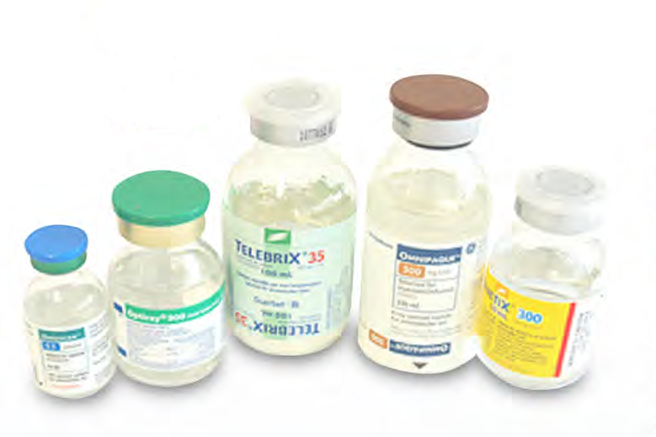
In the past, radiographic diagnostic methods for detecting abnormalities in the large intestine relied on X-ray imaging along with barium enema procedures. Physicians spend considerable time diagnosing abnormalities, which could lead to errors in diagnosis and inappropriate treatment, requiring a significant amount of medication. With the introduction of CT Colonography (also known as Virtual Colonoscopy: VC), it was found that some patients had residual fecal material in the intestines, making it difficult to distinguish between tissue abnormalities and fecal remnants.
Technology readiness level
——-Transfer
——-Prototype
——-Experimental
——-Initial
Technology strengths
Creator
Prof. Dr. Jiraporn Laothamatas
Coordinator: Technology Commercialization